Lecture Details[]
Julian Rood; Week 5 MED1022; Microbiology
Lecture Content[]
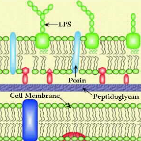
Gram negative cell wall.
Gram negative walls have LPS component of outer membrane; active component is lipid A. This activate complement cascade, stimulate cytokine release, not present in gram positive bacteria as there is no outer membrane. Endotoxins are protein toxins that are secreted, has many effects. LPS structure has an O side chain, core polysaccharide and lipid A which has hydroxyl groups. Loss of O antigen is associated with lack of virulence (mutants are phagocytosed and lysed by serum components). Loss of the core makes the polysaccharide sensitive to hydrophobic compounds such as detergents and bile salts. Lipid A mutants are not viable as they are required for assembly of the outer membrane.
Effects of endotoxin are indirect, host molecules mediate this effect; endotoxin binds to host cell surface and activates LPS pathways. Key cytokines released are TNF-a; IL-1; IL-6. Target cells for endotoxin are phagocytic cells, Kupffer cells in the liver (macrophages surrounding reticuloendothelial system), platelets and granulocytes. It activates the alternative complement pathway and induces various alarm reactions (complement, fever, macrophage stimulation and B cell stimulation). Septic shock is due to high levels of endotoxin. E coli, Pseudomonas aeruginosa and Neisseria meningitidis can cause septic shock. They cause large scale cytokine release, activation of clotting pathways and complement pathways. Systemic effects of shock are fever, shock, blood coagulation, weakness, inflammation and intestinal haemorrhage.
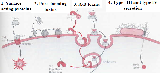
Major classes of endotoxins are those that act directly at target cell surface (a-toxin, perfringolysin O/clostridium perfringens) or those that affect intracellular target molecules (diptheria or cholera toxin). Phospholipases are C perfingens a-toxin; pore forming cytolysins are C perfringens perfringolysin O. A-toxin has phospholipase C activity, hydrolyses sphingomyelin and phosphatidylcholine (components of human cell membranes). A-toxin hydrolyses membrane phospholipids.
Pore forming phospholipids punch holes in the cell membrane and break the permeability barrier, causing death. They can be haemolytic. Large pore-forming toxins bind to cholesterol in the membrane and polymerise forming large pores. Small pore forming toxins are S aureus, leukotoxin. H2O enters the cell causing swelling and lysis.

Toxins that modify intracellular target molecules are most common type of toxin, usually have an A/B structure. A component has enzymic activity, modifies an intracellular target. B component binds toxin to cell membrane and translocates A component into the cell. Simple A-B toxins include diptheria; compound A-B include cholera.
Diptheria is nasopharynx infection, particularly the tonsils. Formation of false membrane leads to excessive inflammation or swelling. Obstructs resp tract, caused by Cornyebacterium diptheriae. Controlled by vaccination to triple antigen. Formed by proteolytic nicking of gene by furin protease and reconstruction. Receptor for the B compound is heparin binding epidermal growth factor-like precursor. A subunit is an ADP-ribosyl transferase, modifies EF2 to inactivate protein synthesis. B moves compound through endosome, A is catalytic, effective component. AB toxin is inserted by lysogenic bacteriophages, which makes it virulent. Diptheria and P aeruginosa target EF2; botulinim and C perfringens iota target actin. Cholera targets Gs proteins.
Cholera is food-borne, Vibrio cholerae. Directly responsible for fluid buildup and diarrhoea. Death is from fluid and electrolyte loss. It is encoded by two separate genes located on genome of filamentous bacteriophage. Nicking occurs outside outer membrane, is assembled in periplasm (between OM and IM), A subunit released in IM. It is endocytosed after binding to a receptor.