Lecture Outline[]
Norm Eizenberg; Week 2 MED1011; Anatomy
Lecture Content[]
Fascia is a fibrous connective tissue
The skeletal system contains 206 bones, 29 in the head, 7 in the neck, 25 in the thorax, 19 in the back, 64 in the upper limbs and 62 in the lower limbs. The axial skeleton contains the CNS and thoracic cage, and the appendicular skeleton makes up the limbs and pelvic girdle.
Bone is a dense connective tissue. It is living with the capacity for growth and remodelling. It contains cells (osteoclasts and blasts) and an extracellular matrix (mineralised ground substance without which would make the bone soft, and collagen fibre reinforcement to protect from being brittle).
Bone contains compact bone, cancellous (spongy) bone with trabeculae, and a medullary cavity. Bony trabeculae are oriented against lines of stress and is both compressive and tensile
Articular surfaces of bones are the only parts not covered by periosteum. The medullary cavity contains red sites for haemopoiesis, and yellow sites which contain fat. Yellow marrow retains the ability to turn into haematopoietic tissue, particularly after blood loss.
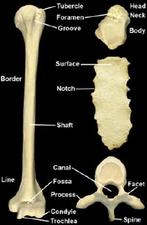
Bones can be long, short, flat, irregular; also pneumatic, sesamoid and accessory. Bony parts and features are articular surfaces at joints, ends and shaft (of long bones), other surfaces, bony features (elevations from fractures), bony markings (grooves from tendons, nerves)
Structures directly related to bone may be damaged in fractures
Cartilage can be hyaline, fibrous (fibrous bundles) or elastic (similar to hyaline but contains lots of elastin); hyaline is avascular and aneural (glassy), unlike cartilage bone requires a blood supply
Bone development can be intramembranous for flat bones of skull or endochondral for all other bones; primary centres occur in 6-8th weeks of embryo and secondary centres appear after birth
Parts of developing bones are the epiphysis (ends of long bone), metaphysis (part of diaphysis that abuts epiphyseal plate), diaphysis (becomes the shaft of a long bone).
Primary centres of ossification appear in the centre of the diaphysis, cartilage is progressively replaced by bone towards the epiphyses. Secondary centres of ossification can be 'pressure' epiphyses at joints or 'traction' epiphyses at attachments of tendons or ligaments. Epiphyseal (growth) plate is a plate of hyaline cartilage responsible for growth in length (occurs at the metaphysial part of an epiphyseal plate). Almost all secondary ossification centres appear after birth (females earlier than males), epiphyseal fusion occurs after puberty (females generally at an earlier age than males). The earlier an epiphysis appears the later it fuses. Epiphyses for long bones tend to appear before (and fuse after) those for smaller long bones.
Growth in width of a long bone is from the periosteum, growth in length occurs more in one end than the other due to nutrient foramen being directed (away from growing end).
Epiphyseal plate may be differentiated from a fracture by comparing with contralateral bone. Epiphyses can be used to predict age as they fuse in sequence. A narrow cleft on a bone indicates an epiphysis undergoing fusion. An injury to epiphysis or epiphyseal plate will impair subsequent growth. Bone infections from spread through the bloodstream tend to occur at the metaphyseal plate. Interruption of blood supply also impairs bone growth.
Blood supply of developing long bones are from the nutrient arteries, periosteal arteries, metaphysial arteries and epiphysial arteries. In development the epiphyseal and metaphyseal arteries are end arteries and do not link with each other, and interruption of either may impair bone growth. Upon closure, they anastomose. Periosteal bones may be extensively stripped during surgery and deprive the underlying compact bone.
A simple fracture stays within the skin, compound fracture breaks out. In adults bones are stronger than ligaments, vice versa in children. Healing of fractures is more rapid in children than adults. Weight bearing bones heal slower than those that don't.